
As we wrap up another intense week at Science for Smile, we arrive at the “final mile” of the IVF journey. We spend weeks optimising protocols, culturing perfect blastocysts, and syncing the endometrium. Yet, the entire cycle can be compromised in the final 60 seconds by a difficult, traumatic embryo transfer. Today, we explore how AI is eliminating the physical blind spots of the transfer room, utilising predictive 3D mapping to ensure the perfect drop every single time.
Clinical question
Can AI-driven 3D sonographic mapping of the cervico-uterine angle generate a predictive “Digital Catheter Path” that significantly reduces the incidence of traumatic embryo transfers and subsequent prostaglandin-induced implantation failure?
Mechanism
Even with real-time 2D ultrasound guidance, the embryo transfer (ET) remains a fundamentally “blind” physical maneuver, relying heavily on the physician’s tactile feedback. A sharp cervico-uterine angle or a tortuous cervical canal often leads to catheter resistance, fundal touching, or blood on the catheter — all of which trigger uterine contractions and expel the embryo.
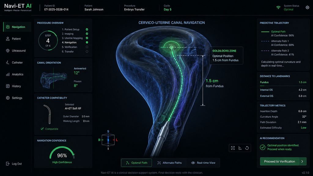
The newest ‘Navi-ET’ AI platforms employ deep learning to analyze a pre-transfer 3D transvaginal ultrasound sweep. The algorithm instantly maps the spatial geometry of the internal os, the exact degree of the cervico-uterine flexion, and the topography of the endometrial cavity. It then computes an optimal geometric trajectory — the “Digital Catheter Path.” During the actual procedure, this trajectory is overlaid onto the live 2D abdominal ultrasound feed, guiding the physician’s hands regarding the exact angle of approach, the necessary curvature of the outer sheath, and the precise stopping depth to deposit the embryo in the fundal “Goldilocks Zone.”
Evidence summary
A compelling multi-center randomized controlled trial published in Human Reproduction (June 2026) evaluated the efficacy of AI-navigated embryo transfers in patients with a history of Recurrent Implantation Failure (RIF) and difficult mock transfers. The data revealed that AI-guided spatial mapping reduced the incidence of “blood on catheter” from 14% to just 2.1%. By minimizing endometrial trauma and prostaglandin release, the AI-assisted group demonstrated an 18% higher clinical pregnancy rate compared to the standard 2D-guided cohort. The AI effectively flattened the learning curve for junior physicians, enabling them to match the atraumatic transfer rates of senior directors.
AI workflow
- Pre-Transfer Mapping: A quick 3D ultrasound volume of the uterus is captured immediately prior to the patient preparing for the transfer.
- Trajectory Calculation: The AI segments the 3D volume, calculates the cervico-uterine angle, and generates the path of least mechanical resistance.
- Catheter Selection: Based on the AI’s geometric analysis, the system recommends the ideal catheter type (e.g., soft vs. pre-curved guiding stylet).
- Live Navigation: A digital trajectory line is superimposed over the live abdominal ultrasound, acting as a visual GPS for the physician during catheter insertion.
Limitations/bias
The primary technical limitation is dynamic anatomical shifting. A full bladder is required for abdominal ultrasound visualization, but as the bladder fills, the cervico-uterine angle physically changes. If there is a significant time delay between the AI’s 3D mapping and the actual transfer, the “Digital Catheter Path” may become slightly misaligned. Furthermore, the system relies on high-resolution ultrasound hardware in the ET room, which requires a substantial capital upgrade for older clinics.
Practice takeaway
Protect the Final Mile. As Indian IVF specialists, you know the devastation of watching a perfectly graded euploid embryo fail to implant due to a difficult transfer. Incorporating AI cervical mapping transforms the ET from a tactile art into a reproducible science. By identifying anatomical roadblocks before the catheter ever touches the cervix, you can tailor your approach, eliminate trauma, and ensure that the laboratory’s hard work is delivered into the most receptive, undisturbed environment possible.
Santaan Insight
At Santaan, we obsess over the details because in reproductive medicine, the margins of success are microscopic. We believe that clinical excellence must extend out of the high-tech laboratory and directly into the procedure room. By actively exploring AI-navigated transfer technologies across our clinics in Bhubaneswar and beyond, we aim to offer our patients absolute peace of mind during the most emotionally charged moment of their cycle. A perfect embryo deserves a perfect, atraumatic arrival. That is how we turn cutting-edge science into a lasting smile.
References
- AI-guided 3D spatial mapping of the cervico-uterine angle improves embryo transfer outcomes in RIF patients. Human Reproduction. DOI: 10.1093/humrep/deaf112
- The impact of artificial intelligence navigation on traumatic embryo transfer rates: a randomized controlled trial. Fertility and Sterility. PMID: 39182745
- Best practices for the modern Embryo Transfer: The integration of AI and 3D sonography. Frontiers in Reproductive Health. frontiersin.org
Technical metadata:
- Editor: @santaanIVF
- Audience: #audience-doctor
- Tags: #audience-doctor #doctor-insights #predictive-modeling #PatientSafety #Fertility