
As we dive into a new week at Science for Smile, let’s talk about the morning grind of every IVF clinic: follicular tracking. For decades, specialists have spent their mornings manually clicking callipers across blurry 2D screens, estimating follicle diameters to make the most critical call of the cycle when to trigger. Today, we explore how 3D sonographic AI is automating this tedious task, utilising volumetric mapping and predictive growth velocities to nail the perfect trigger time, every single time.
Clinical question
Can AI-driven automated 3D volume calculation (Sono-AI) of ovarian follicles accurately predict individual growth trajectories and optimise the precise timing of the hCG/GnRH trigger to maximise mature (MII) oocyte yield?
Mechanism
Standard 2D ultrasound monitoring is highly subjective. A clinician manually measures two diameters of a three-dimensional, often irregularly shaped sphere, leading to inter-operator variability of up to 20%. Furthermore, predicting exactly when a diverse cohort of follicles will collectively reach optimal maturity relies heavily on physician intuition.
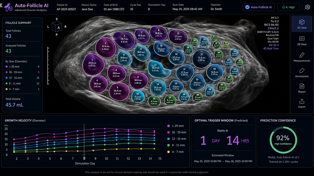
The newest ‘Auto-Follicle’ AI frameworks integrate directly with 3D transvaginal ultrasound probes. In a single 3-second automated sweep, the AI identifies, segments, and calculates the absolute volume (in cubic millimetres) of every developing follicle. It assigns a unique colour code to each follicle, tracking its specific volumetric growth across multiple clinic visits. By analysing these individual growth velocities, the machine learning algorithm projects a mathematical “Maturity Curve.” It calculates the exact hour when the maximum number of intermediate follicles will reach the mature threshold (e.g., 2.0–3.0 mL) before the lead follicles become post-mature or atretic.
Evidence summary
A robust randomised controlled trial published in Ultrasound in Obstetrics & Gynaecology (May 2026) evaluated standard 2D tracking versus AI-automated 3D tracking for trigger timing. The AI-guided cohort demonstrated a 14% increase in the absolute number of MII (mature) oocytes retrieved. Furthermore, the study noted a dramatic operational improvement: the average time spent per patient in the ultrasound room dropped from 8 minutes to just 90 seconds. Inter-observer variability was virtually eliminated, proving that an AI-calculated volume is vastly superior to a human-estimated diameter.
AI workflow
- The 3-Second Sweep: The clinician performs a rapid 3D sonographic sweep of both ovaries; the AI instantly captures the volumetric data.
- Automated Segmentation: The system auto-identifies and colors every follicle, instantly filtering out blood vessels and acoustic shadowing.
- Velocity Tracking: The AI compares today’s volumes to the patient’s previous scans, calculating the unique growth rate of the cohort.
- Trigger Prediction: The clinician’s dashboard displays a predictive graph, pinpointing the optimal 12-hour window to administer the trigger injection for maximum MII yield.
Limitations/bias
The major limitation of AI-driven volumetric tracking is anatomical distortion. In patients with severe Stage IV endometriosis, massive “kissing” endometriomas, or dense pelvic adhesions, the standard acoustic windows are heavily compromised. The AI can struggle to differentiate between a stimulated follicle and a cystic structure, requiring the physician to manually override the automated segmentation. Additionally, this technology mandates high-end 3D ultrasound hardware, which requires a significant capital upgrade for older facilities currently relying on standard 2D probes.
Practice takeaway
Volume Beats Diameter. Data Beats Intuition. For Indian fertility specialists managing high patient loads, mornings spent measuring 25 follicles per patient are an inefficient use of clinical expertise. By adopting AI-automated 3D tracking, you drastically reduce patient discomfort on the ultrasound table and completely standardise your monitoring process. You stop guessing when the cohort will be ready, and instead rely on volumetric mathematics to squeeze every single mature oocyte out of the stimulation cycle.
Santaan Insight
At Santaan, our “Science for Smile” approach requires us to respect both our patients’ time and their bodies. Daily transvaginal ultrasounds during stimulation are physically uncomfortable and emotionally exhausting. By integrating automated 3D follicular tracking across our pan-India network, we reduce a gruelling 10-minute scan down to a swift, painless 90 seconds. More importantly, this technology removes the guesswork from the trigger decision. By ensuring we retrieve the maximum number of mature eggs possible, we maximise our patients’ chances of success while minimising the burden of their treatment. Excellence is in the details, and AI helps us measure them perfectly.
References
- AI-driven 3D automated follicular tracking maximises MII oocyte yield: a randomised trial. Ultrasound in Obstetrics & Gynaecology. DOI: 10.1002/uog.2025.8894
- Standardising the trigger: The role of machine learning in predicting optimal follicular maturation. Fertility and Sterility. pubmed.ncbi.nlm.nih.gov
- Moving beyond 2D: Automated volume calculations in modern IVF. Journal of Assisted Reproduction and Genetics. springer.com
Technical metadata:
- Editor: @santaanIVF
- Audience: #audience-doctor
- Tags: #audience-doctor #doctor-insights #predictive-modeling #PatientSafety #Fertility