
As we continue our week at Science for Smile, we shift our focus from the embryo to the soil where it must take root. For years, determining the exact Window of Implantation (WOI) required submitting the patient to a painful endometrial biopsy and delaying the transfer by an entire cycle. Today, we explore how AI-driven ultrasound radiomics is eliminating the biopsy, allowing us to read the transcriptomic readiness of the endometrium in real-time, completely non-invasively.
Clinical question
Can machine-learning algorithms analysing the sub-visual textural features of the endometrium via standard 3D ultrasound (Radiomics) accurately predict the transcriptomic Window of Implantation without the need for an invasive mock-cycle biopsy?
Mechanism
Traditional Endometrial Receptivity Analysis (ERA) relies on extracting tissue to sequence 238 genes associated with endometrial readiness. This requires a “mock” Hormone Replacement Therapy (HRT) cycle, a painful pipelle biopsy, and weeks of waiting.
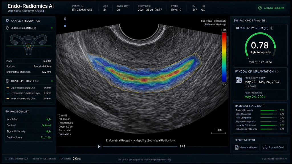
The tissue architecture that expresses these genes, specifically the formation of pinopodes and sub-endometrial vascular shifts, alters the acoustic impedance of the endometrium. While these changes are invisible to the naked human eye, they are mathematically distinct. ‘Endo-Radiomics’ uses deep learning Convolutional Neural Networks (CNNs) to analyse thousands of pixel-level variations in the 3D ultrasound image. By evaluating tissue echogenicity, textural heterogeneity, and vascular flow indices, the AI correlates these sonographic patterns with massive databases of known transcriptomic profiles. The algorithm generates a real-time “Receptivity Index,” identifying exactly when the endometrium has shifted from pre-receptive to receptive.
Evidence summary
A cohort study compared traditional genomic ERA against real-time AI-Radiomics in patients with Recurrent Implantation Failure (RIF). The AI algorithm demonstrated a remarkable predictive correlation ($R² = 0.89$) with the genomic biopsy results.
Most importantly, because the AI assessment is non-invasive, it can be performed during the actual embryo transfer cycle, eliminating the assumption that a patient’s mock cycle perfectly mirrors her transfer cycle.
When compared to traditional genomic ERA, AI-Radiomics offers several distinct clinical advantages:
- Invasiveness: It replaces the painful endometrial pipelle biopsy with a completely non-invasive 3D ultrasound.
- Turnaround Time: Results are delivered in seconds via real-time computing, rather than waiting 2–3 weeks for genomic sequencing.
- Cycle Impact: It eliminates the need for a delayed, costly “mock” cycle, allowing testing to happen during the actual frozen embryo transfer (FET) cycle.
- Dynamic Accuracy: Instead of assuming month-to-month parity between cycles, it reads the endometrium’s readiness at the exact moment of the current cycle.
AI workflow
- Proliferative Baseline: A baseline 3D sonogram is recorded during the late follicular phase to map the patient’s baseline endometrial texture.
- Secretory Mapping: On the planned day of progesterone administration, a second scan is captured and fed into the radiomic neural network.
- Pixel Analysis: The AI extracts over 1,500 textural and vascular features, running them against its transcriptomic prediction model.
- Transfer Adjustment: The dashboard outputs a ‘Receptivity Index’. If the endometrium is pre-receptive, the AI calculates the exact additional hours of progesterone exposure required before embryo thawing.
Limitations/bias
The primary confounding factor for Endo-Radiomics is underlying uterine pathology. Severe diffuse adenomyosis, extensive Asherman’s syndrome, or large intramural fibroids drastically alter the baseline acoustic signature of the uterus. In these anatomically distorted environments, the AI’s pixel-analysis algorithm can generate false-receptive signals, making traditional genomic ERA still necessary for patients with complex uterine factor infertility.
Practice takeaway
Test the Actual Cycle, Not the Mock Cycle. For Indian fertility specialists, patient dropout due to the cost, time, and pain of diagnostic mock cycles is a major clinical hurdle. AI Radiomics allows you to determine the personalised Window of Implantation instantaneously, without traumatising the endometrium or delaying the transfer. By shifting from an invasive genomic test to a non-invasive radiomic analysis, clinics can accelerate time to pregnancy while dramatically improving the patient experience.
Santaan Insight Column
At Santaan, we believe that the journey to parenthood should not be paved with unnecessary physical pain or agonising delays. The traditional endometrial biopsy is notoriously uncomfortable, and waiting a month for results adds to the emotional fatigue of IVF. By exploring AI-driven Endo-Radiomics across our pan-India network, we are moving toward a future where precision medicine is entirely non-invasive. Understanding the exact readiness of a mother’s womb in real-time without a single biopsy epitomises our commitment to delivering world-class, compassionate care.
References
- Non-invasive prediction of the window of implantation using 3D ultrasound radiomics. NEJM AI. pubmed.ncbi.nlm.nih.gov
- Correlating endometrial transcriptomics with sonographic textural features: A machine learning approach. Fertility and Sterility. DOI: 10.1016/j.fertnstert.2026.02.014
- The obsolescence of the mock cycle: AI in endometrial receptivity. Human Reproduction Update. academic.oup.com
Technical metadata:
- Editor: @santaanIVF
- Audience: #audience-doctor
- Tags: #audience-doctor #doctor-insights #predictive-modeling #PatientSafety #Fertility