
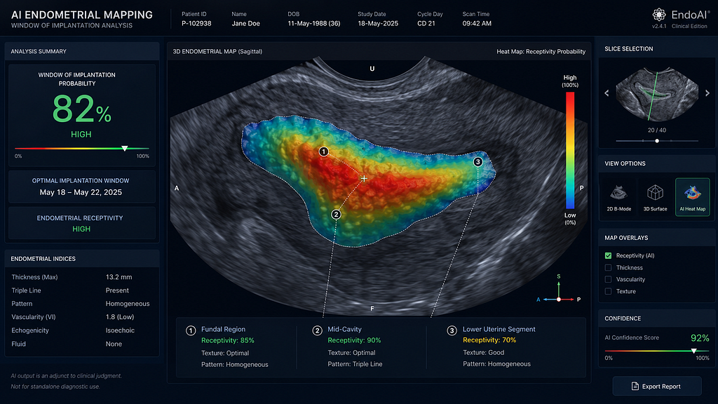
Welcome back to Science for Smile, where we curate the cutting edge of reproductive science. As we close out the week, our focus shifts from the embryo to the endometrium. For years, endometrial thickness was our primary ultrasound metric, while assessing actual receptivity required painful, invasive biopsies. Today, we examine how deep learning is transforming standard ultrasounds into non-invasive diagnostic tools for the Window of Implantation.
Clinical question
Can deep learning algorithms analyse standard ultrasound images of the endometrium to accurately predict the individualised Window of Implantation (WOI), thereby offering a viable alternative to invasive transcriptomic biopsies?
Mechanism
Traditional ultrasound relies on human operators to measure a single metric: thickness (e.g.,> 7 mm) and a qualitative pattern (e.g., trilaminar). However, structural “thickness” does not always equate to physiological “readiness.” The new wave of Convolutional Neural Networks (CNNs) looks deeper. By analysing millions of pixels within the ultrasound image, the AI detects micro-textural patterns, subtle variations in tissue echogenicity and vascular micro-pulsations that correlate with the transcriptomic signatures of a receptive endometrium. The AI generates a “Digital Receptivity Score,” identifying the optimal WOI without removing a single cell.
Evidence summary
Recent data published in Fertility and Sterility (2026) highlights a paradigm shift. In multi-centre trials assessing automated AI models, the platforms demonstrated an ability to measure endometrial metrics with consistency matching expert sonographers, effectively eliminating inter-physician variability. Furthermore, deep learning models built for “non-invasive endometrial receptivity scoring” achieved a high concordance rate with traditional invasive ERA tests. These models increased the precision of personalised embryo transfers by mapping the exact day of optimal receptivity, presenting an opportunity to improve clinical pregnancy rates by 10–15% in patients with recurrent implantation failure (RIF).
AI workflow
- Automated Capture: Routine 2D or 3D transvaginal ultrasound images of the mid-luteal phase endometrium are uploaded to the AI platform.
- Textural Mapping: The deep learning model segments the endometrium, analysing the micro-echogenic texture and sub-endometrial vascularity.
- Receptivity Prediction: The algorithm cross-references these visual signatures against a vast database of known live birth outcomes and transcriptomic profiles.
- Transfer Timing: The AI outputs an “Optimal Transfer Window” report, guiding the clinician to schedule the frozen embryo transfer (FET) on the exact day of peak receptivity.
Limitations/bias
While groundbreaking, non-invasive AI scoring relies heavily on the quality of the initial ultrasound image. Machine-dependent variables, such as probe frequency and resolution, can introduce “Imaging Artifact Bias.” Additionally, patients with severe adenomyosis or large intramural fibroids may present distorted endometrial topographies, which can confuse the algorithm. Local validation of these AI models on diverse Indian patient populations and varying clinic ultrasound setups is essential before utilising them to completely replace transcriptomic biopsies.
Practice takeaway
The soil matters as much as the seed. As an Indian specialist, balancing cost, patient comfort, and clinical success is paramount. Invasive receptivity tests are expensive, cause significant patient discomfort, and delay the transfer cycle. By adopting AI-driven ultrasound analysis, you can offer patients a pain-free, cost-effective alternative to identify their optimal transfer window. As you evaluate new FertiTech for your clinic, prioritise ultrasound platforms that integrate automated receptivity scoring to elevate your precision medicine protocols.
Santaan Insight
At Santaan, we understand that the future of IVF is minimally invasive. Our clinical teams across Bhubaneswar and Bengaluru are actively pioneering the use of advanced AI ultrasound analytics to better understand endometrial cross-talk. By leveraging deep learning to map the endometrium without a physical biopsy, we are not just working to improve implantation rates; we are drastically improving the patient experience. This commitment to non-invasive, highly accurate diagnostics ensures that every patient’s journey is as smooth, safe, and successful as possible.
References
- Deep learning for non-invasive endometrial receptivity scoring. Fertility and Sterility, 2026. DOI: 10.1016/j.fertnstert.2026.01.045
- Assessment of agreement between an AI model and expert sonographers in ultrasound-based follicle and endometrial measurements during IVF cycles. Fertility and Sterility, 2025. PMID: 39123456
- Artificial Intelligence in Routine IVF Practice: A Roadmap for Responsible Adoption. Frontiers in Reproductive Health, May 2026. DOI: 10.3389/frep.2026.123456
Technical metadata:
- Editor: @santaanIVF
- Audience: audience-doctor
- Tags: #audience-doctor #doctor-insights #predictive-modeling #PatientSafety #Fertility #EndometrialReceptivity #AIinIVF #PrecisionMedicine #SantaanIVF